
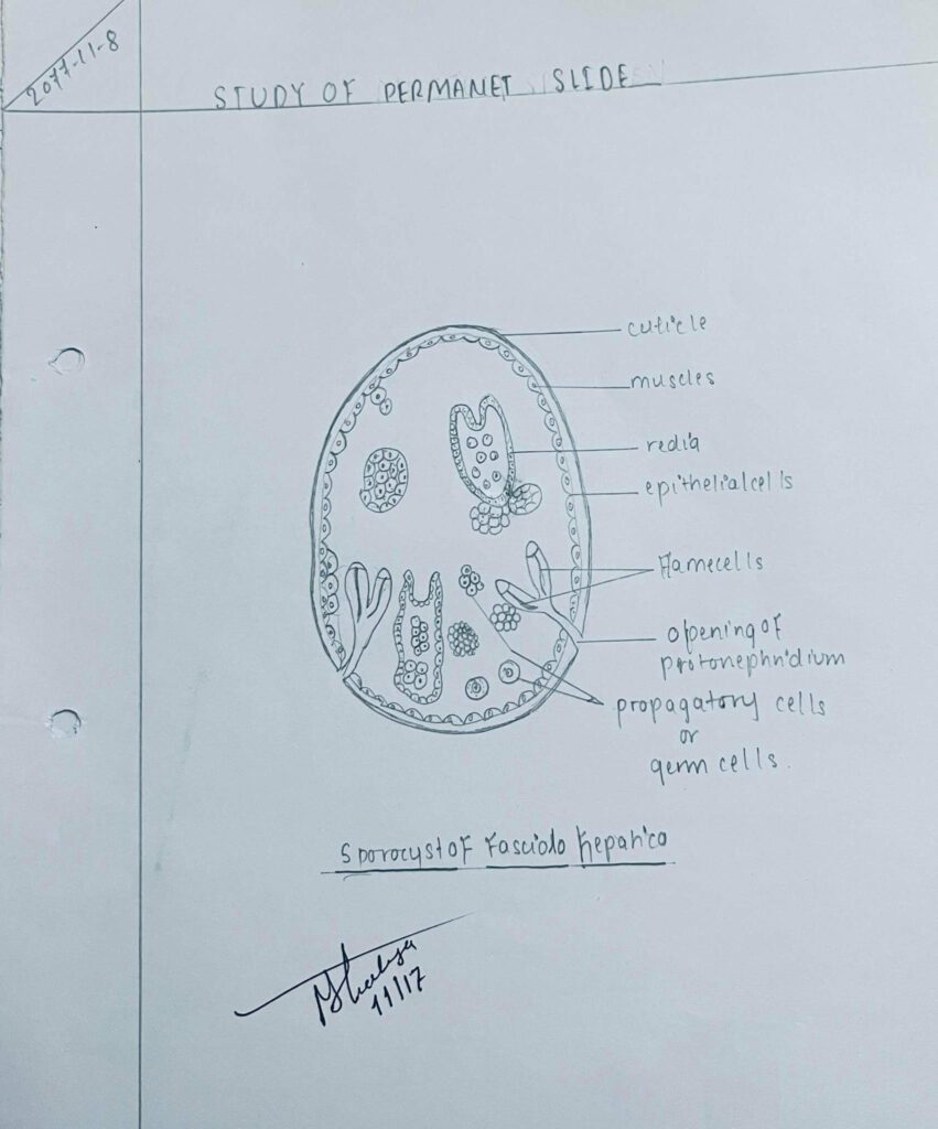
Comment of Sporocyst OF Fasciola Hepatica
1) Sporocyst develops from the miracidium larva in the pulmonary chamber of the snail.
2) The cilia and hexagonal cells covering the body of miracidium are shed.
3) Apical glands, cephalic glands, brain ,eye spots and the primitive guts of miracidium are degenerated.
4) Sporocyst is an elongated sac-like structure covered with cuticles..
5) Body wall consists of sub-epithelial cells, muscles and mesenchyme.
6) Body sac has flame cells and germ cells.
7) Germ cells multiply and give rise to the next larva stage known as redia larva.
Diagram of Sporocyst OF Fasciola Hepatica
Comments of Radia Larva of Fasciola Hepatica
1) Radia larvae develop from the germ cells of the sporocyst.
2) The body of redia is an elongated sac-like.
3) Anterior end bears the mouth leading into the muscular pharynx which finally leads into the sac- like intestine.
4) Just behind the pharynx is a muscular ring-like swelling known as collar, which helps in locomotion.
5) The posterior region is also provided with two stumpy processes known as loppets helpful in locomotion.
6) Just posterior to collar a permanent aperture, containing few germs cells.
7) Germ cells often give rise to second generation daughter rediae.
8) Redia gives rise to a new type of larva known as cercaria larva from the germ cells.
Diagram of Radia Larva of Fasciola Hepatica
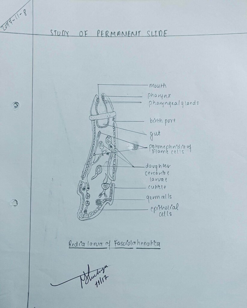
Radia Larva of Fasicola Hepatica