Comments of Enterobius Vermicularis
Distribution:
Enterobius Vermicularis is worldwide in distribution and is specially found in Europe, U.S.A, India, Chile,Africa and canada
Habit and Habitat:
Enterobius Vermicularis is cosmopolitan, but more common in Europe and America. In some communities 40-100 percent of the population may be infected.
General Character:
1. Enterobius Vermicularis is commonly known as pin-worm.
2. It has three small lips and a pair of cephalic expansion at the anterior end.
3. Female is 10mm long with a long pointed tail.
4. Fertilized females make nightly trips to the annus to lay eggs or they may creep out of the anus and lay eggs.
5. Eggs are well advanced when laid, each contains a tad pale like Juvenile.
6. Enterobius causes pin-worm disease, their movement causes intense itching of the anus, inflammation of mucous membrane of colon.
Economic Importance:
It causes enterobiasis in humans.
Diagram of Enterobius Vermicularis
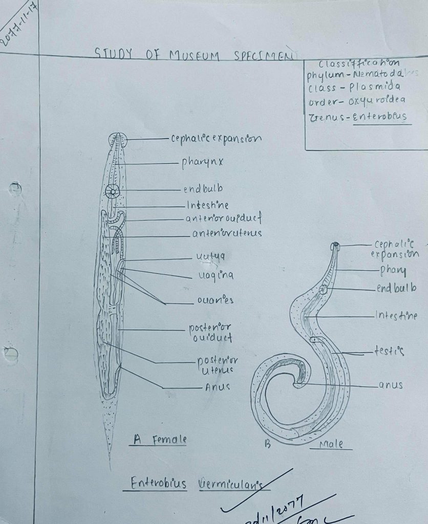
Enterobius vermicularis
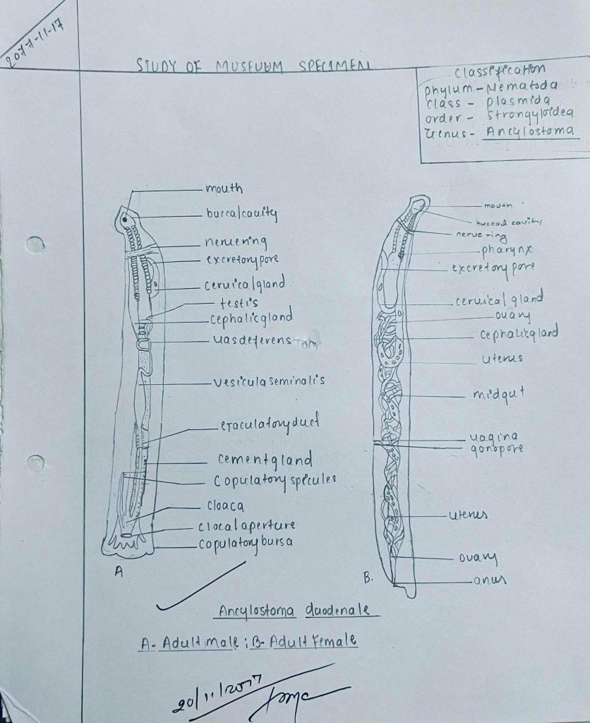
Comments of Ancylostoma Duodenale
Distribution:
Ancylostoma duodenale is endemic in West Europe, tropical and subtropical Asia, Africa and various pacific Islands and South America
Habit and Habitat:
Ancylostoma duodenale is found as an endoparasite in the intestine of man.
General Character:
1. Ancylostoma duodenale is commonly known as horm-worm.
2. Mature worms are cylindrical in shape, narrow anteriorly and white, gray in color.
3 Males measure 8-11 mm in length and females measure 10-13 mm in length.
4. Buccal cavity is oval and the buccal capsule is made of articulated grooved portion.
5. Male is provided with a campanulate bursa which is broader than long and supported by fleshy rays.
6. Vulcca of the female is behind the middle of the body and the tail is tipped by a minute spine.
Economic importance:
It causes ancylostomiasis disease in humans.
Diagram of Ancylostoma Duodenale