
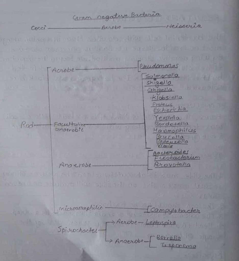
Introduction:
In 1884, a Danish Bacteriologist Hans Christian Gram developed a gram staining technique. Gram staining is the differential staining technique. This technique differentiate bacteria into two major groups as Gram positive and Gram negative bacteria. In this process, bacterial Smear is made by following staining reagents as the primary stain.
i) Crystal Violet:
Gram positive bacteria retain the crystal violet and hence appear deep violet in colour. Gram’s Iodine serves as mordant a substance that forms an insoluble complex by binding to primary stain. The resultant crystal Violet – Iodine (CV-I) complex serves to intensify the colour of stain and all cells appear purple black at this point.
ii) Decolorizing agent:
Ethyl Alcohol(95%) serves as a dual function as liquid solvent and as protein dehydrating agent. Gram negative bacteria contain a higher percentage of lipid than gram positive bacteria. During staining gram negative bacteria alcohol treatment intereact’s lipids which result in increased porosity or cell wall. Thus CV-I complexes can be extracted and gram negative bacteria are decolorized. But gram +ve bacteria, due to their low lipid contents become dehydrated during alcohol treatment. The spore size which decreases in CV-I complex can’t be extracted so they remain purple in colour.
iii) Counter Stain:
Safranin is used for counterstain. Since Gram negative bacterial cells undergo decolorization and absorb counterstain. Hence, gram negative bacteria loses CV colour and counterstained by safranin and appear as red in colour Likewise, gram +ye bacteria retain crystal violet and hence appear red in colour.
Materials Required:
i) Bunsen Burner
ii) Inoculating loop
iii) Glass slide
iv) Bibulous paper
v) Microscope
vi) Gram’s Iodine
vii) Crystal Violet
viii) 95% ethyl alcohol
ix) Safranin
x) Glass rod
Procedure:
i) A clean and grease free slide was taken.
ii) A loopful of water was taken in the slide by a sterilized loop.
iii) A loopful of bacterial suspension was transferred to the slide by the help of a glass rod.
iv) The smear was air dried and heat fixation was done.
v) Then the smear was flooded with crystal violet and left it for one minute and then washed.
vi) After that Gram’s Iodine was added to the smear and waited for 30 seconds and then washed.
vii) Then ethyl alcohol was passed drop by drop to smear for 20 sec. and washed immediately with water.
viii) Finally, safranin was added to the smear and left for 1 minute.
ix) After the smear, was washed with water and then the slide was air dried with bibulous paper and was examined under oil immersion.
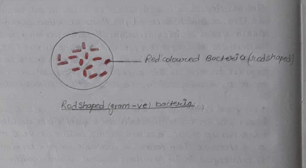
Observation Table:
| S.N | Staining technique | Staining reagent used | Colour of bacteria | Shape of bacteria | result |
| 1 | Gram staining | Crystal violet iodine ,alcohol safranin | Red in colour | Rod shaped | Single arrangement of rod shaped red bacteria was observed |
Result:
The organism was blue violet in colour. Hence, Rod shaped red bacteria was seen.
Conclusion:
So, in the above experiment, bacterial cells which retain crystal violet appear deep violet in colour and are stained by counter Stain. Safranin appear red in colour and are gram negative bacteria. In gram negative, high lipid concentration is found in the outer layer of the cell wall. In gram positive, small amount of lipid content in cell wall is readily dissolved by the action of Alcohol causes formation of minute cell wall pores. So, the primary stain is difficult to remove and cells remain purple. Thus by applying staining dyes in the smear we performed that it was gram negative bacteria.
Precautions:
i) The culture must be fresh that is not older than 24 hours.
ii) Outer delocalization should not be done as it may result in a loop of primary stain.
iii) The culture should be fresh as gram +ve cells tend to lose their ability to retain primary stain.
iv) It must be imperative that slides be thoroughly washed under running tap water or distilled water between application of reagents.
References:
Shah PK; Dahal PR, Amatya J. 2013 “Practical Microbiology” revised edition Delta offset press; Thopathali Ktm Page No.33-38