THEORY:
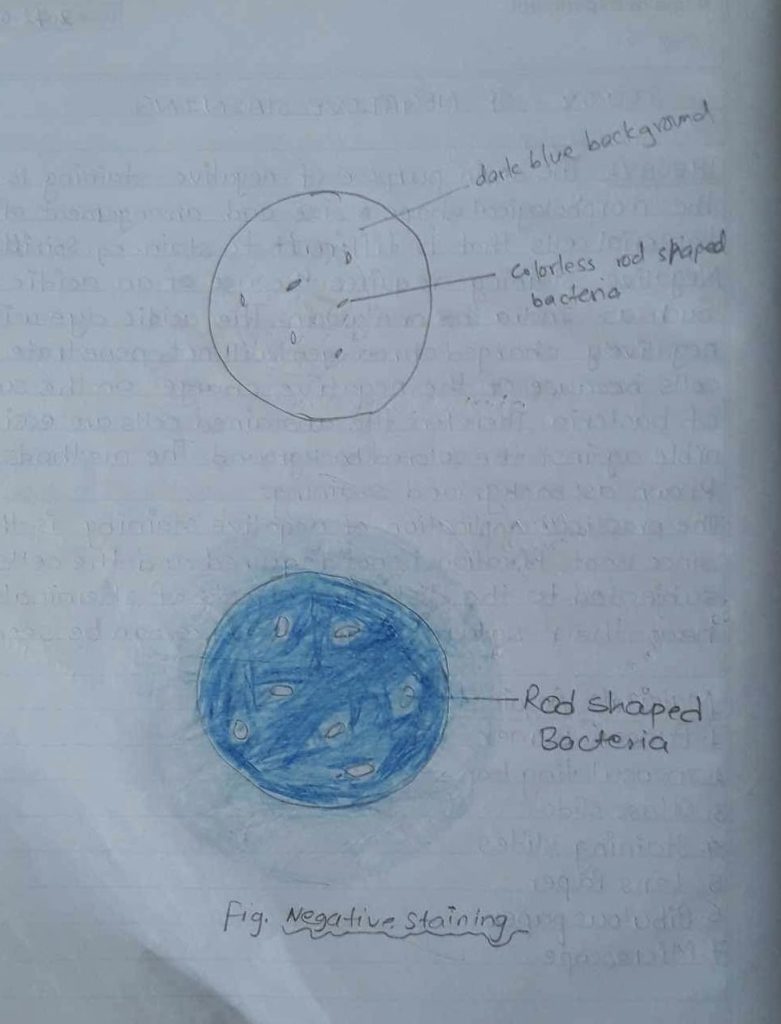
The main purpose of negative staining is to study the morphological shape, size and arrangement of the bacterial cells that is difficult to stain. eg. Spirit Negative staining requires the use of an acidic dye. Such as India ink or nigrosin. The acidic dye with its negatively charged chromogen, will not penetrate the cells because of the negative charge on the surface of bacteria. Therefore, the unstained cells are easily discernible against the colored background. The method is also known as background staining.
The practical application of negative staining is that, since heat fixation is not required and the cells are not subjected to the distorting effects of chemicals and heat, their natural shape and size can be seen.
Materials Required:
1. Bunsen Burner
2. Inoculating loop
3. Glass slides
4. Staining slides
5. Lens Paper
6. Bibulous paper
7.Microscope
Reagent: Indian Ink or Nigrosin.
Culture: 24 hours agar slant culture of Bacillus Cereus micrococcus luteus.

PROCEDURE:
1) The slide was cleaned properly.
2) A small drop of nigrosin was placed from one end of the slide.
3) Aseptically, one loopful of inoculum was placed from the given culture in the drop of nigrosin.
4) A thin smear was made with the help of another slide (held at 30° angle) by sliding over the slide containing the sample.
5) The slide was dried in air but avoided heat fix.
6) At blast the prepared slide was examined under the microscope 1st in low power and then in oil immersion.
OBSERVATION:
| S.N | Staining method | Stain used | Color of organism | Shape of organism | Result |
| 1. | Negative staining | Nigrosin (Acidic) | colourless | Rod shaped | Rod shaped transparent organism was observed |
RESULT AND DISCUSSION:
The bacterial cells with colourless chain in arrangement and rod in shape i.e. Bacillus cereve was observed. By using a negative staining method, we found the organism to be colourless having rod shaped which was defected by the stain nigrosin. The background was black/blue coloured.
CONCLUSION:
Hence, the negative staining of bacterial cells (E . Coli) was done in laboratory with the help of acidic dye i.e. Nigrosin.
PRECAUTIONS:
1) The slide must be absolutely clean: the presence of any residual grease or dirt on the slide will produce an uneven smear.
2) The loop should not be too hot..
3) The amount of nigrosene or India ink should drop.
4) Heat fixation is not necessary as heat destroy the shape of organism.
Reference:
Shah PK, Dahal P.R and Amatya J., Practical microbiology [Revised edition] April 2009]. Delta offset press, thapathali Kathmandu PageNo. 33-38
