
Introduction:-
Microscope is an instrument that magnifies minute objects which are invisible to our naked eyes. It is an important apparatus for microbiology laboratories. Since most of the micro-organisms refer to Unicellular Microscope helps to study them and their Morphological Structure as well. The magnification attainable by microscope ranges from 100x to 400000x. There are several types of microscope available and many techniques have been developed by which we can easily study the micro-organisms.
Types:
Basically Microscopes are of two categories.
i) Light (Optical) Microscope
ii) Election Microscope
i)Light microscope:
It is the type of microscope in which Magnification is obtained by a system of lenses using light waves. It includes following types of microscope
a) Dark Field microscope:The effect produced by dark field technique is that of a dark background against which objects are brilliantly illuminated.
b) Bright Field Microscope: In bright field microscope, Microscopic (area observed) is brightly lighted and organisms appear dark because they absorb some of the light.
c) Phase Contrast Microscope: It is extremely valuable for studying living unstained cells and is widely used in applied and theoretical, biological studies. It is a conventional light microscope fitted with a phase contrast condenser.
d) Fluorescence Microscope: This microscope is used in many diagnostic procedures for identifying microorganisms by using fluorescent staining. It gives a magnification of 1500X and resolution of 100-200 mm.
ii) Electron Microscope:
The electron microscope as the name suggest, was a beam of electron in place of light waves to produce the image specimens can be examined either by :
a) Transmission or
b) Scanning electron microscope
a) Transmission Electron microscope (TEM): It is used to view the Ultrastructure of micro-organisms including viruses. It has much greater resolving power, (1-2) nm and useful magnification (5,00,000) – (1,000,000) X that can be achieved with a light microscope.
b) Scanning Electron microscope (SEM): It is used for showing the detailed structure of the micro-organism.
The normal student’s microscope is a compound light microscope working under the principle of bright field microscope.
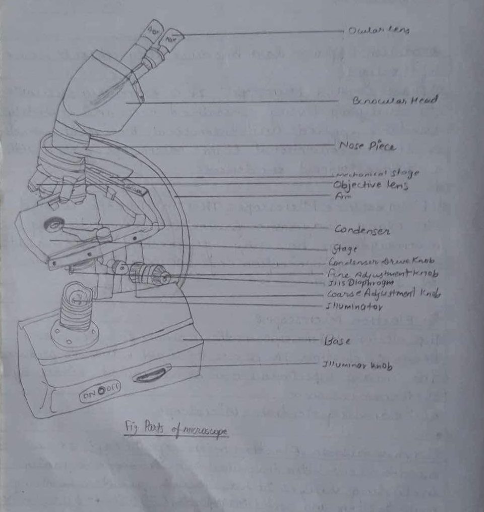
Parts of Microscope:
The components of a microscope that can be broadly categorized into two parts.
A. Mechanical Parts
B. Optical parts.
A. Mechanical Parts:-
The metal structures of the microscope that are used for support purpose are the mechanical parts these includes:
i) Base or foot: The lowermost part which supports the instrument.
ii) Arm or limbs: The U-shaped structure hinged to the foot at the inclination point.
iii) stage: The part situated below the arm where the object which is to be studied is placed. It bears the slide clips and field hanging knobs.
iv) Body Tube: The structure above the arm which holds the lens. It can be moved up and down by
rotating following two knobs.
a) Fine adjustment Knob: A pair of small knobs. It is used to focus Specimen in high magnification.
b) Coarse Adjustment Knob: It is used to focus specimens in loco magnification.
B. Optical parts:-
The optical parts lying below the stage are the accessories while those contained in the
body-tube are essential which are directly involved in magnification. These includes:
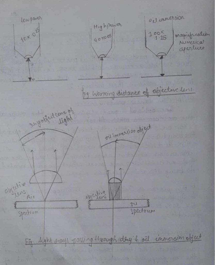
i) Objective Lenses:- These are situated below the tube (i.e. the power part of the body tube). They are of Magnifying power (10x, 20x,40x and 100x) namely, low power objective and oil
immersion objective respectively. Those lenses are mounted on a revolving nosepiece which can be rotated to bring the desired lens in place.
ii) Eyepiece or Ocular lens: The upper end of the body to hold the eyepiece which may be one of the 5X, 10X, or 16x magnifying power which are replaceable.
Magnification:
It means simply the increase in size of the image of an object. The total magnification is the product of the magnifying power of the objective and that of the eye piece.
Magnification table:
| Objective | Eye piece | magnification | Used for viewing |
| 10x | 10x | 100x | Bacterial smear |
| 40x | 10x | 400x | Fungi and protozoa |
| 100x | 10x | 1000x | bacteria(needalimmersion) |
Numerical Aperture:
The numerical aperture (NA) can be defined as a function of the diameter of the objective lens in relation to its focal length.
NA= nSinθ
where ‘n’ is the refractive index of the medium filling the space between lens and object (n=1 for air and n = 1.65 for oil immersion)
Resolving Power:
The smallest distance at which two points can be seen separately is called resolving power of lens.
Resolving Power = wavelength of light/2 x Numerical aperture
= λ /2NA
or, Resolving limit (d) = 0.5λ/nSinθ
= 0.5λ /NA
Where, λ is the wavelength of light source employed.
θ is half angle of light system passing from objective to lens system
n is the refractive index of medium.
Low Power 10x
For 10x
Total angle = 64°
θ=32
NA = nSin32 = 1 xSin32 = 0.52
Resolving Power = λ/2XNA (Bluelight is best)
= 6.8/2×0.52
= 6.8mm/1.04
=6.538μm
High dry 40x
Total angle = 96
θ=48 degree
Numerical aperture =n x Sin48
= 1 x 0.74
= 0.74
Resolving Power (R.P) = λ/2xnSinθ
=λ/2xNA
= 0.73mm/2X1Sin48
= 0.73/2×0.74
= 0.5μm
High Wet (100x)
Total angle = 116
Total angle = 116
θ= 58
N·A=nSin 58
= 1.65x Sin58 [ n = 1.65 for 1oox powered lens)
= 1.399
Now,Resolving Power (R.P.)= λ/2xN.A
= 0.12/2X1.399
= 0.05μm
= 0.12/0.79
= 0.15μm
Note: λ For 10x = 6.8mm.
λ for 40x=0.73 mm
λ for 100x = 0.12 mm
Conclusion:
Hence the microscope used in laboratory was studied and its application in modern microbiology.
Precautions:
i) The microscope must be kept free of dust and moisture.
ii) The lens must be touched by pointers not be hand.
iii) Lens paper or list free optically safe tissue should be used on the lens surface.
Reference:
Shah PK, Dahal PR, Amatya Jyoti, (2009), “Techniques in microbiology” Practical book. Delta offset Press.