COMMENTS ON PINUS.
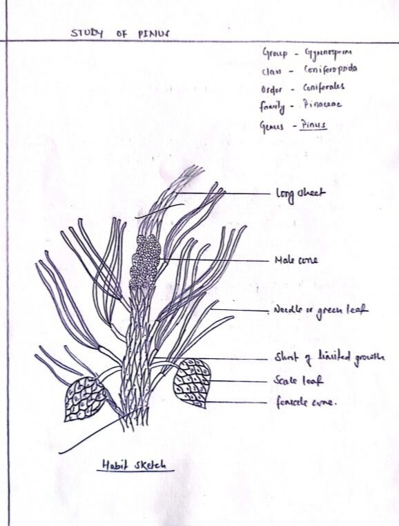
Habit
Pinus is a large resinous gigantic tree widely distributed in the northern hemisphere in temperate regions.
Morphological structure
i) Root:
The primary root is a typical tap root with strong and widely spread lateral roots that are covered with ectotrophic mycorrhiza.
ii) Stem:
The plant has a stout, tall and erect cylindrical stem covered with woody scaly bark. It has regular and symmetrical branches arising from the axial scale leaves.
iii) Leaves:
The leaf is dimorphic, characterized by two kinds of leaves: the scale leaves and foliage leaves. The shoot of limited and unlimited growth bears brown membranous scale leaves. The dwarf shoots give out circular green long needle-like foliage leaves.
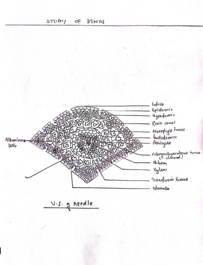
Internal structure of the leaf
i) Cuticular Layer:
The layer is externally coated by a highly cutinized outer layer of epidermis.
ii) Epidermis:
It is the outermost layer composed of tightly arranged small calls interrupted by depressions of Sunken stomata.
iii) Hypodermal layer:
Just beneath the epidermal layer lies one or two layers of sclerenchyma hypodermal layer which is again interrupted by a sub-stomatal air-chamber of sunken stomata.
iv) Mesophyll layer:
The underlying Mesophyll region contains a large number of their walled chlorenchyma tous cells with numerous peg- like infolding of walls that project into the cell cavity.
v) Endodermis:
The Mesophyll cells end at the central circular endodermal layer composed of thick walled borrel shaped compactly arranged starch containing cells.
vi) Pericycle:
Just beneath the endodermis lies a multilayered pericycle of thin cell walled parenchymatous cells.
vii) Vascular bundles:
There are two vascular bundles placed centrally facing phloem bundles towards the upper flat surface of leaf and xylem bundles towards the convex lower surface.
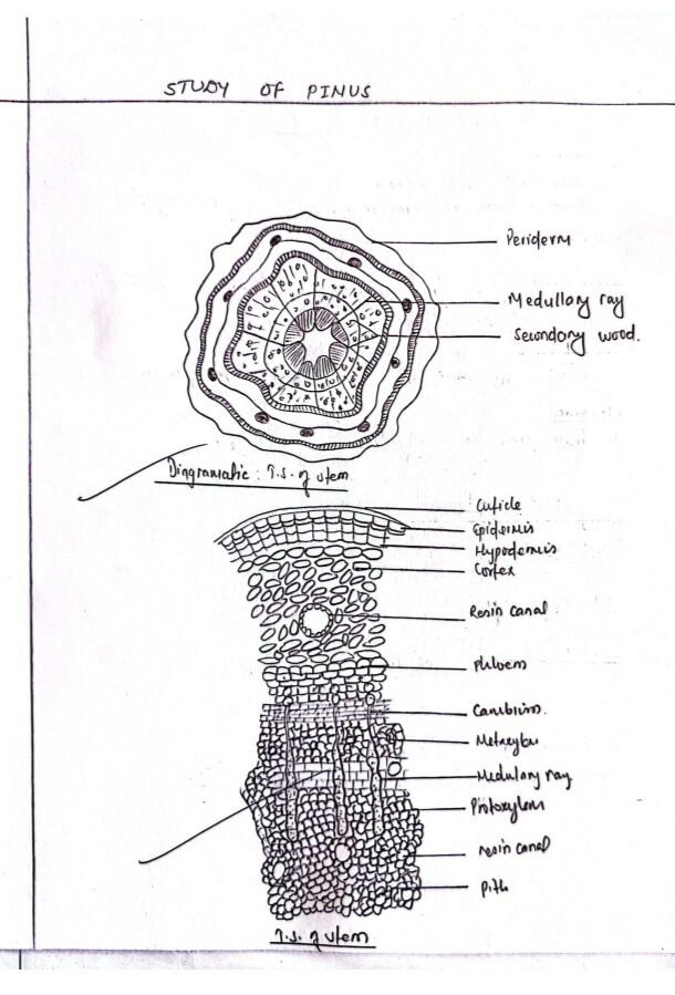
Internal structure of stem
i) Epidermis:
The outermost covering layer is composed of compactly packed one layered cell, whose external wall is highly cutinized.
ii) Cortex:
The epidermis is followed by a few layers of cortex divisible into upper sclerenchymatous hypodermis and lower parenchymatous mass enclosing a number of resin-canals.
iii) Endodermis:
It is the single layered innermost cell ring of the cortex.
iv) Pericycle:
An inconspicuous layer below the endodermis.
v) Vascular bundles:
Many vascular bundles are arranged in a ring. They are conjoint, collateral and open.
vi) Medullary rays:
The vascular bundles are separated by long parenchymatous medullary rays connecting Cortex with pith.
vii) Pith:
The Central space is occupied by parenchymatous pith cells, some of which are filled with tannin.
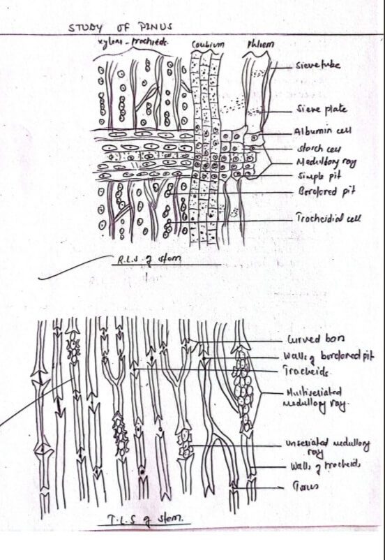
Radial longitudinal section of Stem
i) Secondary xylem:
The tracheids with bordered pits are the characteristic feature of pine. The elongated cells are arranged regularly on radial rows.
ii) Secondary phloem
The sieve tube consists of elongated tapering cells which bears siene plates on the radial walls.
iii) Secondary Medullary rays:
Medullary rays are much more regularly arranged in secondary xylem.
iv) Marginal ray-tracheids:
The upper and lower Margins of medullary rays are accompanied by horizontally seen one-two rows of marginal ray tracheids.
Tangential longitudinal section of stem.
i) Bordered pits:
Above and below the pit, the middle lamella is thickened to form a cured bar at the upper and lower side enclosing clearly a visible torus at the center of it.
ii) Uniseriate Medullary rays:
There are uniseriate medullary rays with only one cell broad and less than a dozen of cells in height.
iii) Multi-seriated medullary rays:
Some medullary rays are multiseriate with more than one called broad.
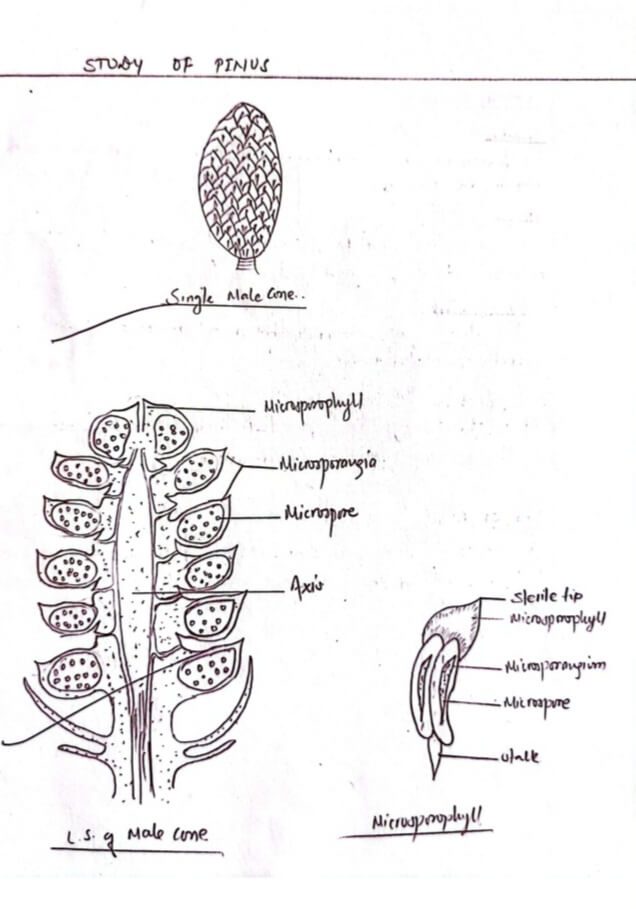
Male cone
i) Position:
The Male cone is small Measuring about 1/2 to 1 inch in length. It develops in clusters.
ii) Shape:
A single cone is composed of a central elongated axis surrounded by 60-100 spirally arranged scales leaves.
iii) Microsporophyll:
Each microsporophyll is flattened and triangular in outline with slightly upwardly bent sterile up and a short stalk below.
iv) Microsporangium:
The ventral surface of microsporophyll possesses two swollen but elongated pollen-sacs called microsporangia.
v) Microspores:
The microspore is a small tri-layered unicellular structure having external incomplete cutinized exine, middle complete exo-intire and inner thin intire.
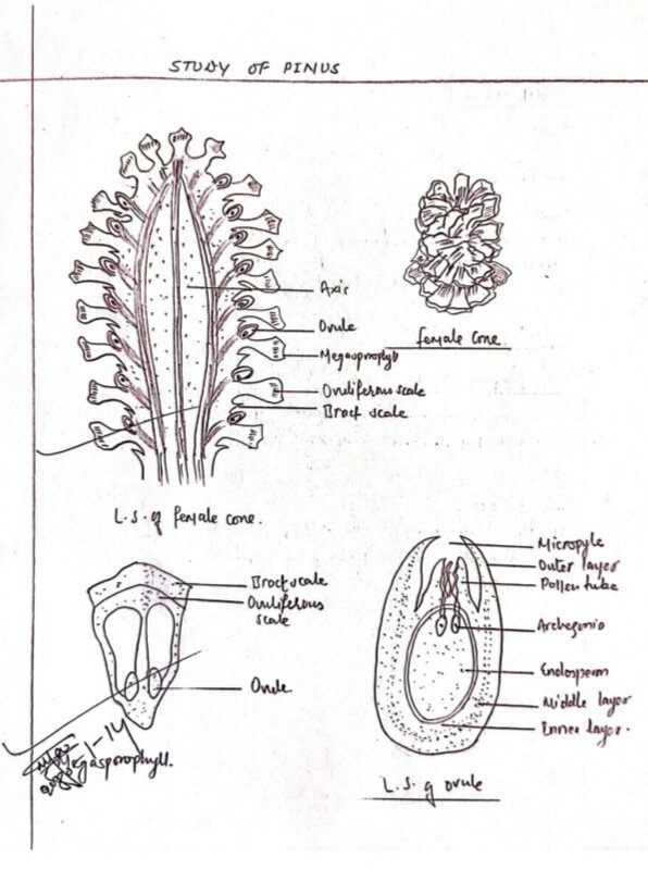
Female cone
i) Position:
The female cone is a prominent structure borne in clusters of 1-4 in the axil of scale leaves of branches of unlimited growth.
ii) Shape:
The cone consists of central axis bearing spirally arranged megasporophylls composed of two types of bracts.
a) Bract scale:- It is the outer smaller leathery bract scale developed directly from the axis.
b) Ovuliferous scale:- It is the triangular expanded bigger woody scale borne on the upper surface bract scale.
L.S. of Ovule
The ovule consists of a central mass of tissue called nucellus fully enclosed by three layered integuments: outer and inner fleshy and middle stony layer. The integument remains fused with nucellus but separated from a short distance near the micropyle. A few archegonia lie at the apex of the nutritive endosperm formed within the nucleus.