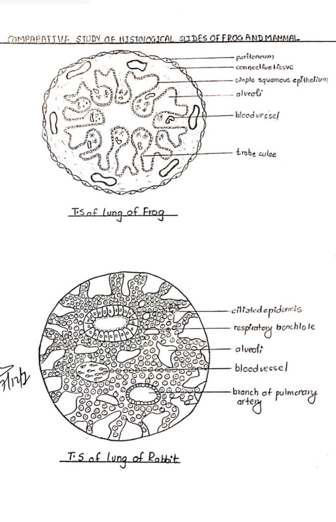
Comments on T.S of lung of Frog
i) The outer wall of the lung is peritoneum consisting of connective tissue which contains some elastic fibers.
ii) The peritoneum is covered externally by squamous epithelium or mesothelium.
iii) The central cavity of the lung is partly divided into numerous chambers or alveoli separated from each other, partitions or trabeculae.
iv) The trabeculae are lined partly by a thin, flattened simple squamous epithelium and partly by a ciliated columnar epithelium on the outer edges.
v) The walls of trabeculae are richly supplied with blood vessels and capillaries.
vi) Numerous bundles of muscle fibers are present within the trabeculae.
Comments on T.S of lung of Rabbit.
i) It consists of numerous alveoli.
ii) The alveoli communicate with one another by apertures in their wall.
iii) Around each alveolus is a network of capillary blood vessels in connection with the pulmonary artery or vein of the lung.
iv) Numerous alveoli form clusters which open in an alveolar duct.
v) Each bronchus as it enters the lungs, divide and subdivide into finer and finer branches, the bronchioles.
vi) The bronchioles are divided into respiratory bronchioles.
vii) The respiratory bronchiole gives rise to several alveolar ducts which open into alveoli or air-sac.